Heart disease is the leading cause of death worldwide, and heart transplantation is currently the only treatment available for patients with heart failure. Parallel to this, however, there is also a problem related to donation, where there is a severe shortage. These are also part of the reasons for the attempts to develop new approaches to regenerate the diseased heart.

By utilizing a bioink that contains components of contractile organs and stem cells from individual patients, Harvard University's Wyss Institute research team has developed the ability to 3D print heart tissue that closely resembles the structure of the human heart muscle. This breakthrough innovation presents an exciting opportunity for scientists to produce thicker, multilayered muscle tissue with more realistic physiological contractile characteristics, ultimately paving the way for the creation of heart tissue replacements using 3D printing technology.

Dr. Jennifer Lewis, a key contributor to the research at the Wyss Institute, emphasizes the critical importance of accurately replicating the contractile system of the heart at every level, from individual cells to thicker, multilayered tissue, in order to successfully generate functional heart tissue for use in replacement therapy.

Harvard, Wyss, and Trestle Biotherapeutics

For several years, the Wyss Institute has been at the forefront of developing groundbreaking bioprinting technologies and tissue engineering techniques. In February 2022, Trestle Biotherapeutics, a bioengineering startup, was able to secure a license for a technology developed by Harvard University, through the Wyss Institute, that enables the speedy production of kidney tissues.

Researchers from Harvard University are actively involved in the bioprinting of heart tissue, having developed a specialized 3D printing technology in 2018 specifically for this purpose. The goal of their work is to examine the efficacy of artificial heart valves. One year later, in 2019, they introduced the SWIFT technology, which enables the bio-ink printing of contractile components derived from human organs.

In simple terms, the technology can be described as follows: the team uses pre-assembled contractile elements created from human induced pluripotent stem cells - cells created in the laboratory that have the properties of embryonic cells. These cells have the ability to produce vascular networks using bioink. In this way, scientists have created cardiac tissue constructs that have the typical high cell density of normal heart tissue.
Harvard 3D printed tissue / 3D принтирана тъкан от Харвард
3D printed heart tissue

Medthodology and Results

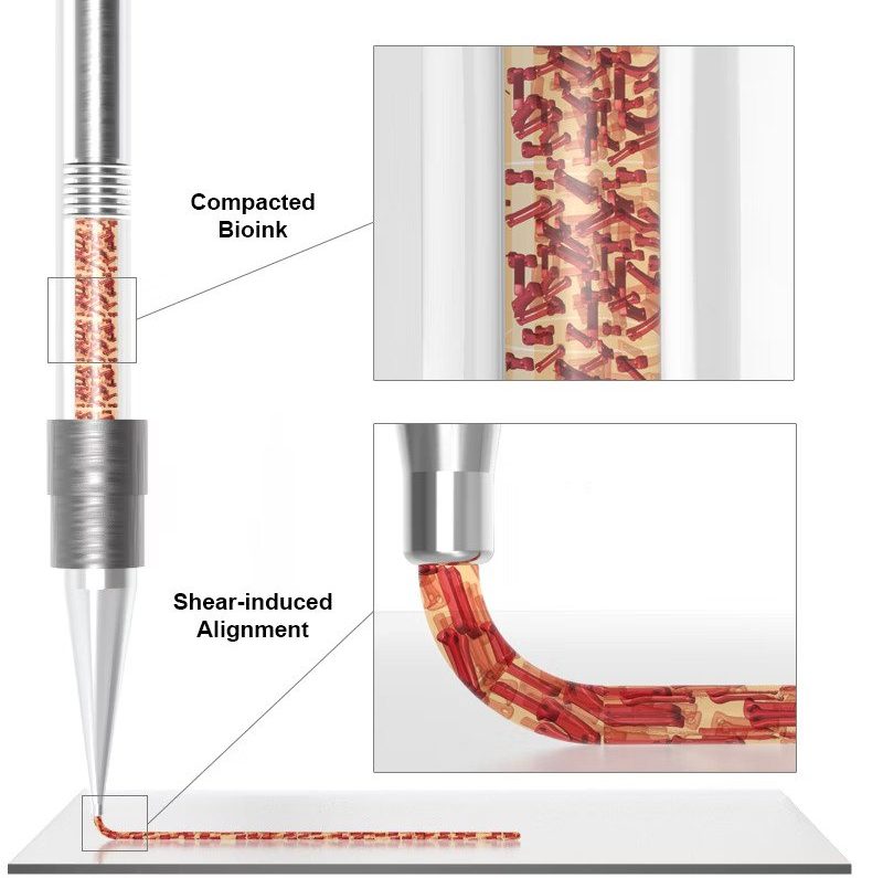
In order to create a bioink rich in cells, the research team combined fibroblast cells, which make up a significant portion of the body's connective tissue, with extracellular protein collagen (ECM). As the cells compact the collagen, a dense microtissue is formed. These contractile organ blocks can then be used as raw materials for the bioprinting process.

The team observed the deformations of the printed microfilaments and discovered that tissue contractile forces and shrinkage rates increased over the course of seven days, meaning that the 3D printed tissues matured and developed into actual muscle fibers.

The selection of engineering materials used in bioprinting of human organs or components is a crucial consideration as it can impact their compatibility with the body and the risk of implant rejection.

The ideal biomaterial for bioprinting human organs or parts should possess similar biochemical and mechanical properties as the patient's own tissue. Furthermore, for bioprinted heart tissues to be effective, they must be fully compatible with the patient's immunological, cellular, biochemical, and anatomical properties.

Moving forward, the Harvard research team intends to expand their method to create more complex, multilayered tissues. The ultimate goal in this field is to successfully generate a fully functional heart that can be implanted.

Currently, 3D-printed biotissue can be utilized to create myocardial patches that can replace damaged tissue after a heart attack, similar to LEGO bricks. This technology can also be applied to newborns with congenital heart defects.

For More News from the World of 3D Printing...

Visit our blog regularly and follow us on Facebook. If you have your own ideas related to 3D printing and prototyping, don't hesitate to reach out to our expert team today.
© 2022 SolidFill, All Rights Reserved
44 "Zhitnitsa" str., 1612, Npz Stedets, Sofia, Bulgaria
+359 888 722 894
© 2022 SolidFill, All Rights Reserved
linkedin facebook pinterest youtube rss twitter instagram facebook-blank rss-blank linkedin-blank pinterest youtube twitter instagram